.
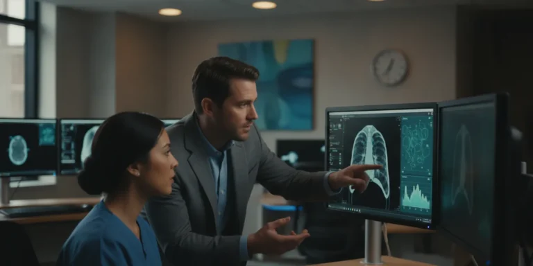
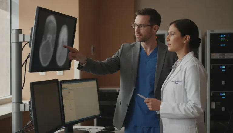
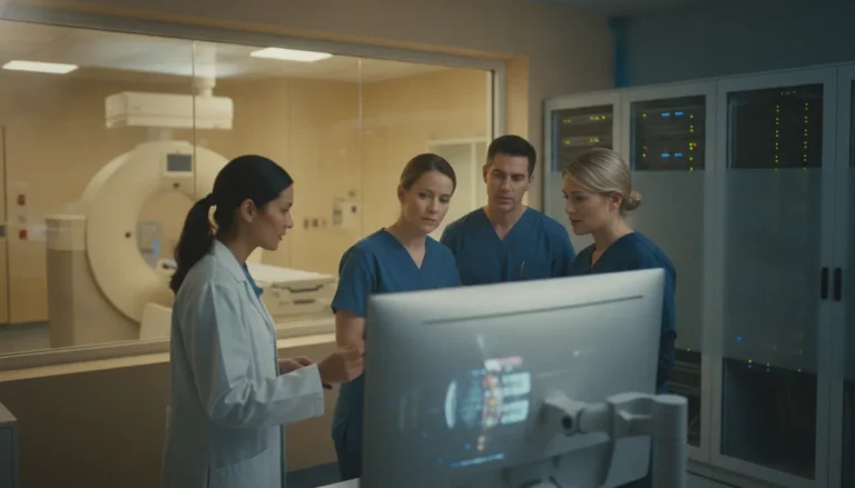
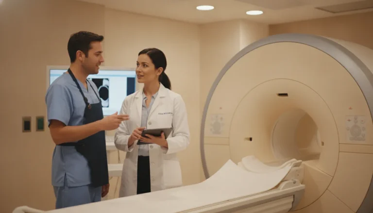
Medical imaging does not end when a scan is captured. A study must move reliably from the modality to a standards-based record. It then passes through storage and retrieval, diagnostic review, and specialist collaboration. Gaps at any point can create avoidable friction for radiology, IT, and referring teams.
The lifecycle of medical images typically includes acquisition, DICOM transmission, PACS storage, quality and diagnostic review, secure exchange, and specialist collaboration. A connected workflow preserves the study and its relevant information while applying access controls, encryption, and auditability appropriate for protected health information.
Understanding what happens at each stage makes it easier to evaluate interoperability, governance, and clinical workflow fit. It also clarifies why real-time modality streaming during an examination complements, rather than replaces, the completed DICOM study.
What Are Medical Images, and Why Does the Workflow Matter?
Medical images are digital representations produced by imaging systems such as computed tomography (CT), magnetic resonance imaging (MRI), X-ray, ultrasound, mammography, fluoroscopy, and PET/CT. In a healthcare environment, they are more than visual files. Each study must remain connected to the information needed to identify, retrieve, review, interpret, report, and share it appropriately.
The Digital Imaging and Communications in Medicine (DICOM) standard provides the common framework for transmitting, storing, retrieving, processing, and displaying medical imaging information. A DICOM object pairs image data with related study and patient information, helping systems preserve the context required for authorized review. This standardization is foundational to interoperability across modalities, radiology information systems, picture archiving and communication systems (PACS), viewers, and connected clinical applications. Learn more about the DICOM standard.
The medical-image lifecycle
A dependable workflow follows the study from its point of origin through each operational handoff:
- Acquisition: An imaging modality captures the study and generates the associated data.
- Quality review: Staff confirm that the study is complete, properly associated, and suitable for the next step in the workflow.
- Transmission and storage: DICOM data moves to an archive, commonly a PACS or cloud-based imaging environment, where it can be governed and retrieved.
- Retrieval and diagnostic review: Authorized clinicians access the relevant current and prior studies through an appropriate viewer. Diagnostic interpretation remains the responsibility of qualified clinical professionals.
- Reporting and exchange: Findings and images move to the systems and people involved in the patient’s care, referral, consultation, or operational review.
The National Institute of Biomedical Imaging and Bioengineering describes medical-imaging workflows in terms that include acquisition, quality review, storage, retrieval, interpretation, reporting, and exchange. That full sequence matters because a technically excellent image can still create operational friction if it is difficult to locate. Separated from its metadata, restricted by fragmented access, or unavailable to the specialist who needs to review it. See the NIBIB overview of medical imaging and data resources.
For radiology directors and IT leaders, workflow design therefore extends beyond selecting a viewer. It includes modality connectivity, DICOM routing, archive strategy, identity and access controls, auditability, interoperability, and secure collaboration across sites. A platform should support the organization’s existing architecture while giving authorized teams a consistent path from acquisition to review and exchange. Teleray’s radiology solutions reflect this broader operational view, connecting medical imaging workflows with collaboration needs while keeping the clinical team in control of interpretation.
How Are Medical Images Captured and Encoded in DICOM?
The path from an examination to a usable imaging study begins at the modality. CT, MRI, X-ray, ultrasound, mammography, fluoroscopy, and PET/CT systems generate image data in different ways, but the operational goal is consistent: capture the study. Associate it with the correct patient and examination, review its quality, and transmit it in a format that downstream systems can understand.
DICOM, or Digital Imaging and Communications in Medicine, is the international standard for transmitting, storing, retrieving, processing, and displaying medical imaging information. It is more than an image file. A DICOM object pairs the pixel or frame data with related information needed to identify and interpret the study, including study and series context. That structure lets imaging systems exchange information without treating every exam as an isolated picture.
Acquire the study at the modality
The technologist performs the examination using the appropriate modality and protocol. A CT or MRI study may contain multiple image series, while ultrasound and fluoroscopy can involve moving images or captured frames. Mammography, X-ray, and PET/CT produce their own modality-specific data and presentation requirements. The acquisition system records the source data and begins associating it with the scheduled examination.
Associate the images with the correct study
Patient and order information must remain connected to the images as the study moves through the imaging workflow. Modality Worklist integration can provide scheduled exam details to the modality, reducing manual re-entry and supporting consistent identifiers. The DICOM metadata travels with the image data so receiving systems can organize the exam by patient, accession, study, and series.
Review image quality and completeness
Before transmission is treated as complete, the imaging team reviews whether the required views, series, or cine captures are present and usable for the intended examination. This is a workflow quality check, not an automated guarantee of diagnostic adequacy. Clinical professionals remain responsible for assessing the study and determining whether additional acquisition is needed.
Encode and transmit the DICOM objects
The modality packages the image data and associated metadata as DICOM objects. Then sends them through a configured connection to a destination such as a PACS, cloud archive, or diagnostic workstation. Standards-based DICOM services support functions such as image storage, routing, and query and retrieval. TeleRay supports DICOM 3.0, standard transfer syntaxes, DICOM routing, and Query/Retrieve functions including C-FIND, C-MOVE, and C-GET. It also supports direct modality connections through DICOM Store SCP/SCU and related integration methods.
Prepare the study for authorized downstream use
After transmission, the receiving system validates and indexes the study for storage, retrieval, review, or exchange. Where appropriate, de-identification tools can remove or transform identifying information for approved uses. Access controls and governance are essential because DICOM metadata may contain protected health information. The completed DICOM study can then support diagnostic review, specialist collaboration, or secure sharing without losing the context attached at acquisition.
Live modality streaming is a complementary workflow rather than a replacement for this process. During a procedure, real-time viewing can support remote collaboration, while the completed DICOM files remain available for storage, retrieval, and later review.
For a standards-based overview of DICOM information and services, see the DICOM standard.
Where Do Medical Images Go After Acquisition? PACS, Cloud Storage, and Retrieval
After acquisition, medical images need more than a place to sit. They must remain associated with the correct study, available to authorized users, and accessible in a form that supports review and collaboration. In most imaging environments, the workflow centers on DICOM, the standard used to transmit, store, retrieve, process, and display medical imaging information. A DICOM object includes both image data and related information needed to identify and interpret the study.
A picture archiving and communication system, or PACS, manages the archive and retrieval layer. It receives studies from imaging modalities, stores them with their associated data, and makes them available to approved users through a diagnostic viewer or connected clinical workflow. Retrieval is therefore not simply a file search. It depends on accurate patient and study information, appropriate permissions, and systems that can exchange imaging data without losing the context required for review.
For a deeper explanation of the archive and retrieval process, see how PACS archives and retrieves medical images. Healthcare organizations should also distinguish archival storage from image exchange. Sharing a study with an outside specialist, facility, or care team may require a separate secure workflow, even when both parties use PACS.
| Approach | Where the archive is managed | Key considerations |
|---|---|---|
| Local PACS | On infrastructure operated or controlled by the healthcare organization | Requires planning for hardware, storage capacity, access, maintenance, backup, and continuity. Connectivity to outside reviewers must be designed deliberately. |
| Cloud PACS | In a hosted environment accessed through a network connection | Can support distributed access and reduce the need to operate all archive infrastructure locally. Review the provider’s security controls, availability commitments, data governance, and integration model. |
| Hybrid PACS | Across local and hosted environments | May accommodate existing systems and phased modernization. It requires clear rules for synchronization, ownership, retention, access, and the authoritative copy of each study. |
The right architecture depends on the organization’s existing modalities, network design, clinical workflows, governance requirements, and collaboration model. Regardless of where the archive resides, medical images can contain protected health information. Access controls, encryption, auditability, and appropriate agreements are therefore part of the storage decision, not optional add-ons. A secure DICOM exchange and storage workflow can help organizations extend access beyond a single facility while preserving control over who can view and share each study. Learn more about secure DICOM image exchange and storage.
How Are Medical Images Reviewed and Shared Securely?
Secure review begins with a controlled diagnostic environment, not simply a larger screen or a faster file transfer. Medical images may contain protected health information, so healthcare organizations need safeguards that govern who can open a study. What they can do with it, and how activity is recorded. The U.S. Department of Health and Human Services Security Rule addresses administrative, physical, and technical safeguards for electronic protected health information. In practice, access policy must be designed alongside the imaging workflow.
A diagnostic viewer should support the clinical task without obscuring the study’s context. Authorized users may need to compare prior examinations, review multiple modalities, apply measurements, or use specialty tools while preserving the original DICOM study. TeleRay’s diagnostic viewer is FDA 510(k)-cleared for primary diagnostic interpretation. That is a specific regulatory clearance for the device’s intended use, not an endorsement of a particular diagnosis or a substitute for the judgment of a qualified clinician. For a practical evaluation framework, see these DICOM viewer considerations.
Access should follow role and responsibility
Role-based access control helps align permissions with job responsibilities. A radiologist may require diagnostic tools, while a referring clinician may need access to a report and selected images. An imaging technologist may need to manage acquisition and routing without receiving the same privileges as an interpreting physician. Mandatory multi-factor authentication adds another verification step, while documented provisioning and deprovisioning processes reduce the risk of former or unauthorized users retaining access.
Encryption protects information as it moves and while it is stored. TeleRay documents AES-256 encryption at rest, TLS 1.3 in transit, and proprietary peer-to-peer encryption described as exceeding AES-256. Its platform also documents isolated storage silos for each customer, geo-redundant storage, and Azure infrastructure. These controls should be assessed as part of a broader security program, including business associate agreements, incident response, retention rules, and organizational governance.
Auditability supports accountable exchange
Secure sharing requires more than sending a link. The system should make it possible to control recipients, limit access, and maintain an audit trail of relevant events, such as viewing, downloading, routing, and sharing. Standards-based DICOM exchange can preserve interoperability across imaging systems, while de-identification tools can support approved teaching, research, or collaboration use cases when patient identity is not required.
TeleRay combines DICOM image exchange with cloud PACS and supports multi-site study sharing. That can help teams establish a repeatable process for referrals and consultations rather than relying on ad hoc media or email attachments. Read more about diagnostic image sharing and the governance decisions that protect study integrity. HIPAA compliance and SOC 2 Type II are part of TeleRay’s documented compliance framework. Organizations should still validate configuration, contracts, user roles, and retention practices against their own policies and applicable requirements.
What Is the Difference Between Image Sharing and Live Modality Streaming?
Image sharing and live modality streaming support different points in the imaging workflow. The distinction matters because a completed study and a procedure in progress create different operational requirements for radiology teams, referring clinicians, and remote specialists.
Post-exam image sharing begins after a modality has captured the study. The resulting DICOM files can be transferred to a PACS, retrieved for authorized review, or exchanged with another organization. The receiving team is working with a completed set of medical images. They can examine the study, compare it with prior exams when available, document an interpretation, and communicate findings through the appropriate clinical workflow.
This model is essential for routine diagnostic access and consultation. It creates a durable record that can be stored, retrieved, and shared according to organizational policies. A secure DICOM exchange and cloud PACS can help connect sites without requiring every participant to be in the imaging room at the time of acquisition. For a deeper look at the live workflow, see live radiology observation and modality streaming.
Live modality streaming addresses a different moment: the scan or procedure is still taking place. Rather than waiting for the completed study to arrive, a remote specialist can observe the imaging process in real time. Teleray Live supports real-time streaming from modalities such as CT, MRI, and ultrasound, along with live video conferencing. Depending on the configured workflow, participants may also see the room and probe positioning, which provides context that a static image alone cannot provide.
That context can support collaboration while the local team is acquiring the study. A remote specialist may observe the modality output, discuss the workflow with on-site staff, and provide guidance within the responsibilities and governance defined by the healthcare organization. The technology does not replace the local team, clinical judgment, or the formal diagnostic interpretation process. Instead, it adds a communication and observation layer to the procedure.
The two workflows can also operate together. After the live session ends, the complete DICOM study can be transferred for storage, review, and documentation. In this sequence, streaming supports real-time collaboration, while DICOM exchange and PACS provide the persistent medical record for subsequent access. Treating them as complementary capabilities helps healthcare leaders evaluate whether a platform supports both immediate specialist access and reliable post-exam management.
When comparing solutions, ask whether the system only moves completed files or can also connect authorized specialists to the procedure as it happens. That answer affects equipment connectivity, user permissions, audiovisual design, and integration planning. It also clarifies whether a proposed platform addresses image distribution alone or the broader workflow around acquisition, collaboration, and diagnostic review.
How Can Specialists Collaborate Around Medical Images?
Specialist collaboration depends on more than sending a file from one inbox to another. A useful referral workflow preserves the study, its identifying context, the reason for consultation, and the discussion that follows. That continuity helps each authorized participant work from the same clinical record while keeping responsibilities clear.
Start with a complete referral context
A referring team may need to share a DICOM study with a radiologist, surgeon, emergency physician, or another specialist. The referral should travel with the relevant patient and encounter information, the imaging modality, the acquisition date, the referring question, and any prior studies that inform the review. DICOM objects are designed to pair image data with related information needed to identify and interpret a study, but the surrounding workflow still matters.
Organizations should define who can initiate a consultation, who can view the study, and how comments or reports are returned to the referring team. Role-based access and auditable activity are especially important when medical images contain protected health information. The goal is not simply broad access. It is controlled access for the right people at the right point in the referral process.
Connect PACS, viewers, and the EMR
When a specialist must leave the PACS, diagnostic viewer, and EMR or EHR to reconstruct the case, essential context can become fragmented. A connected workflow can make it easier to locate the study, open it in an appropriate viewer. And return the consultation record to the system used by the care team. Interoperability should be evaluated at the level of actual workflows, including patient matching, study retrieval, report exchange, and permissions.
Teleray reports support for more than 250 EMR and EHR systems. That documented capability is a platform feature, not a guarantee that every organization will have the same implementation. IT and clinical leaders should confirm the specific systems, interfaces, data elements, and governance requirements involved. See the EMR integration resources when assessing how connected clinical workflows may fit an existing environment.
Support consultation without losing the imaging record
Secure DICOM exchange and cloud-based access can support referrals across facilities, including organizations that do not share the same local infrastructure. A specialist can review the authorized study, communicate relevant observations through the established consultation process, and leave the original record available for appropriate follow-up. This is different from live modality streaming. During a procedure, live streaming can provide real-time visibility into CT, MRI, or ultrasound activity. After the exam, the completed DICOM study remains the durable record for storage, retrieval, and diagnostic review.
For teams designing this model, radiology imaging workflows provide a useful starting point for evaluating modality support, diagnostic viewing, exchange, and specialist access together. The platform should fit the organization’s clinical governance, security controls, and established review responsibilities rather than attempting to replace them.
What Should Healthcare Leaders Evaluate in a Medical Imaging Platform?
A credible evaluation should look beyond the image viewer. Healthcare leaders need to understand how medical images move through the organization. Who can access them, and whether the platform fits the clinical and technical environment already in place.
Interoperability and workflow fit
Start with standards and connections. Ask whether the platform supports DICOM 3.0, standard transfer syntaxes, routing, Query/Retrieve, and Modality Worklist. These capabilities determine how studies move from scanners into storage and downstream systems. Confirm how the platform connects with the organization’s PACS, radiology information system, and electronic medical record. Including whether it supports the modalities in use, such as CT, MRI, ultrasound, mammography, or PET/CT. TeleRay reports integrations with more than 250 EMR and EHR systems, but each deployment still requires a workflow-specific technical review. EMR integration should be assessed in the context of actual users, sites, and data flows.
Security, governance, and resilience
Medical imaging studies may contain protected health information, so evaluate more than a security statement. Look for encryption in transit and at rest, mandatory multifactor authentication, role-based access, audit trails, customer isolation, retention controls, and defined administrative responsibilities. Confirm where data is stored, how backups are protected, and how the organization would recover access after an outage. TeleRay’s documented environment includes Azure infrastructure, isolated storage silos for each customer, geo-redundant storage, AES-256 encryption at rest, TLS 1.3 in transit, and role-based access controls. Its compliance materials specify HIPAA compliance and SOC 2 Type II. Those designations should be reviewed alongside the organization’s business associate agreement, policies, and risk assessment.
Diagnostic capability and collaboration
Determine whether the viewer supports the intended clinical use. TeleRay’s diagnostic viewer has FDA 510(k) clearance for primary diagnostic interpretation and includes tools such as multiplanar reconstruction, maximum intensity projection, 3D volume rendering, fusion imaging, and measurements. This is a precise clearance statement, not an implication of FDA endorsement. Validate performance with representative studies and specialty workflows, while keeping clinical judgment with qualified professionals.
Finally, assess scalability and collaboration. Look for multi-site sharing, prior-study comparison, disaster recovery, and controls that remain manageable as users and facilities grow. Separate ordinary post-exam DICOM exchange from real-time collaboration: TeleRay Live supports live streaming from CT, MRI, or ultrasound during procedures, alongside video, room context, and later DICOM transfer. A platform earns consideration when these capabilities work together without forcing teams into disconnected tools.
Frequently Asked Questions
What is the difference between DICOM and PACS?
DICOM is the standard that defines how imaging information is formatted, transmitted, stored, and displayed. PACS is the system that archives and retrieves imaging studies for authorized review. In practice, DICOM enables interoperability, while PACS organizes access to the resulting studies.
How are medical images protected when they are shared?
Because imaging studies may contain protected health information, secure sharing should include role-based access, encryption, multifactor authentication, and auditability. Healthcare organizations should also confirm that vendors support appropriate HIPAA safeguards and data-governance agreements.
Can specialists view an imaging procedure in real time?
Yes, live modality streaming can provide a real-time view during a CT, MRI, or ultrasound procedure, allowing a remote specialist to collaborate with the onsite team. This is different from sharing the completed DICOM study after the exam.
What should a healthcare organization look for in a diagnostic imaging platform?
Evaluate DICOM interoperability, PACS retrieval, diagnostic viewer capabilities, secure exchange, EMR connectivity, access controls, and support for the modalities used across your sites. If primary diagnostic interpretation is part of the workflow, verify the viewer’s regulatory status. Teleray’s viewer is FDA 510(k)-cleared for primary diagnostic interpretation, according to its compliance documentation.
Schedule a Demo of Your Medical Imaging Workflow
Understanding how medical images move across acquisition, storage, review, and collaboration can clarify where a unified platform may fit your organization. Teleray can help your team examine these workflows, including secure exchange and live modality streaming, in the context of your operational needs. To discuss the platform with our team, Schedule a demo.