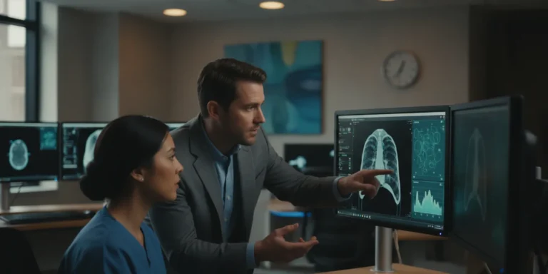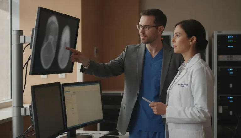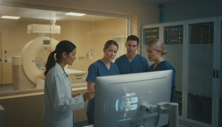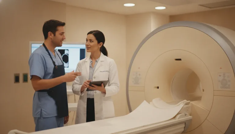

Medical imaging workflows depend on more than the scanner itself. A health system also needs a reliable way to capture, organize, store, retrieve, and share diagnostic images without forcing clinicians to manage disconnected systems or physical media.
A PACS, or picture archiving and communication system, is the imaging infrastructure that moves studies from modalities such as CT. MRI, and ultrasound into secure digital archives and makes them available to authorized users. It typically works with DICOM, the standard format for medical images, and connects with clinical workflows so radiology teams can review and distribute studies efficiently.
Understanding how these systems work helps radiology directors, imaging managers, and IT leaders evaluate storage, viewing, security, interoperability, and emerging capabilities. The foundation is the system’s role in making diagnostic imaging accessible across the organization.
What Is a PACS and Why It Matters
A Picture Archiving and Communication System, or PACS, is the digital infrastructure used to acquire, store, transmit, and display medical images. It connects imaging modalities with secure storage and clinical workstations so authorized teams can retrieve and review studies without relying on physical film. PACS is therefore more than an image archive. It is a core operating layer for modern radiology and hospital-wide digital imaging.
The transition from film to digital workflows changed how radiology departments manage studies and collaborate with other clinical services. A review indexed by PubMed describes PACS as the technology that enabled radiology’s move from analog film-based operations to digital workflows. In practice, that shift reduces dependence on physical handling and makes imaging data available through a controlled electronic environment.
For radiology directors, the value is operational consistency. Images from multiple modalities can be stored economically and accessed from a central system rather than maintained in separate physical locations. CT, MRI, ultrasound, radiography, and other supported studies can be managed within a common imaging infrastructure, subject to the organization’s modality, storage, and viewing requirements. This gives radiologists and referring clinicians a more practical way to locate the relevant study and review it as part of an established workflow.
For imaging center managers, PACS also supports scale. As study volume grows, a digital archive can provide a more sustainable foundation than film-based storage and manual retrieval. A quantitative evaluation of a filmless radiology department reported gains in workflow efficiency and quality compared with traditional processes, including stronger coordination across the hospital environment. The findings are available through PubMed Central.
PACS matters at the enterprise level because imaging is no longer confined to the radiology reading room. It must be available to the departments, specialists, and care teams that legitimately need it, while remaining protected as sensitive patient data. Research on hospital-wide PACS adoption has examined its role as essential infrastructure for clinical users across a health system. Reinforcing that PACS should be evaluated as a strategic platform rather than a standalone storage purchase. The system’s architecture, interoperability, security controls, and support for clinical workflows all deserve attention during selection.
The Core Components of a PACS
A PACS is an operating architecture, not simply a place to save images. Its four main components are imaging modalities, a secured network, clinical workstations, and archives. Together, they move a study from acquisition to review while maintaining the organization and controls required for sensitive patient information. The architecture is commonly described as the foundation of digital medical imaging workflows.
| Component | Role in the PACS workflow |
|---|---|
| Imaging modalities | Acquire the study and generate image data (CT, MRI, ultrasound, radiography, mammography). |
| Secured network | Transport images between modalities, servers, and workstations with access and privacy controls. |
| Clinical workstations | Let radiologists and clinicians retrieve, view, and compare studies from one interface. |
| Archives and central storage | Preserve studies for retrieval, review, and consultation across the health system. |
1. Imaging modalities
The workflow begins at the modality. CT, MRI, ultrasound, radiography, mammography, and other systems acquire the study and generate the image data. The modality supplies the clinical source data, along with identifying and study-level information needed for downstream management. A PACS then receives that data in a standardized form, allowing images from different manufacturers and departments to enter a consistent workflow.
2. A secured network
The network connects modalities, servers, workstations, and authorized users. After capture, images travel across this controlled infrastructure rather than being moved as physical film or isolated files. Network design affects availability, responsiveness, access controls, and the ability to share studies across locations. Because imaging data is protected health information, security and privacy safeguards are essential, including controls that support HIPAA obligations.
3. Clinical workstations
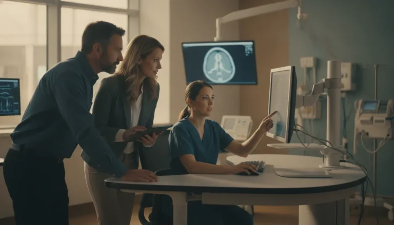
Workstations are where radiologists and other authorized clinicians retrieve and review studies. A well-designed PACS can provide convenient access to images from multiple modalities through a single workstation, reducing the need to search across separate systems. The workstation should support the viewing and comparison tasks required by the clinical team, while permissions ensure that users see only the information appropriate to their role.
4. Archives and central storage
The archive preserves studies so they can be retrieved for current review, comparison with prior exams, or consultation by another authorized specialist. In the standard workflow, PACS captures images from a modality, stores them on a central server, and distributes them through the network for clinical review. Storage strategy must account for volume, retention, redundancy, backup, and controlled access. Organizations evaluating ultra-secure cloud PACS storage should assess these safeguards alongside performance and interoperability.
When these components work together, the result is a continuous path from image capture to clinical interpretation. The modality creates the study, the secured network transports it, the archive keeps it available, and the workstation presents it to the right reviewer. That coordination is what turns separate imaging devices into a usable enterprise imaging workflow.
How a PACS Captures and Stores Images with DICOM
A PACS connects imaging modalities to a digital workflow. When a CT, MRI, ultrasound, or other modality completes an examination, the system sends the resulting images through the clinical network to a central archive. This capture, storage, and distribution sequence is the basic operating model of PACS: images are acquired from modalities. Stored on a central server, and made available across the network for review. This workflow is described in more detail here.
DICOM, or Digital Imaging and Communications in Medicine, provides the common standard that allows imaging equipment, archives, and viewing applications to exchange medical images. It is the universal format used for PACS image storage and transfer, rather than a proprietary file type tied to one manufacturer. That standardization is important in environments where a health system operates equipment from multiple vendors. The images can move through the same archive and viewing workflow without requiring staff to manage separate storage methods for each modality.
From modality to central archive
After acquisition, the PACS receives the study and stores it electronically on central servers. The archive becomes the organized source for current examinations and prior imaging, subject to the facility’s retention, access, and security policies. Digital storage removes the physical handling requirements associated with film and supports a more consistent way to manage imaging data. A medical imaging review describes PACS as a technology for digital acquisition, archiving, transmission, and display, reflecting the shift from analog film operations to digital radiology workflows: PubMed review of PACS history and development.
DICOM also helps keep studies associated with the appropriate patient and examination context as they move between systems. In practical terms, that means staff can search for an examination, retrieve the relevant image set. And compare it with available prior studies through the PACS interface instead of locating film or navigating disconnected modality-specific archives. The system’s value is not simply that it stores large image files. It creates a consistent pathway for finding and reviewing studies.
Access from one workstation
Once stored, images from multiple modalities can be accessed from a single workstation. A radiologist or other authorized clinical user can retrieve a study when needed. Review the images digitally, and access relevant imaging history without moving between physical files or separate viewing stations. This convenient access is one of the defining practical advantages of PACS, particularly for organizations managing imaging across departments or locations. Effective implementation still depends on appropriate network capacity, archive management, user permissions, and security controls, but DICOM provides the shared technical language that makes the workflow possible.
How Radiologists Access and Share Images Across a Health System
Once images are stored in a PACS. Authorized radiologists and referring clinicians can retrieve them through a connected workstation rather than locating physical film or moving records between departments. A single workstation can provide convenient access to studies from multiple imaging modalities, giving the care team a more complete view of the patient’s available imaging history.
This access model reduces the manual work associated with image management. Radiologists can review relevant studies in real time, spend less time searching for prior images, and focus more of their attention on interpretation. Because images are maintained digitally, the workflow also reduces the risk of a physical study being misplaced or lost before it reaches the clinician who needs it.
Sharing studies with specialists
PACS supports image sharing among specialists across departments and locations. A radiologist can make a study available for review by the appropriate referring clinician or another specialist without relying on duplicate film copies or physical transport. This is particularly important when a case requires input from more than one clinical team. Faster access to the same image set can support more timely diagnostic decision-making, while preserving a consistent digital record for the professionals involved.
For radiology directors, the operational value is broader than faster image retrieval. Digital access helps connect radiology with the rest of the health system. Allowing imaging information to move through established clinical workflows instead of remaining isolated in the imaging department. It can also reduce avoidable delays caused by missing film, incomplete handoffs, or repeated requests for the same study.
Supporting patient throughput
When images become available sooner and can be reviewed without manual handling, radiology teams can process studies more efficiently. Research on radiology workflow and patient volume has examined the effect of PACS on technologists and radiologists, with digital image availability associated with improved patient throughput. The practical goal is not to accelerate clinical judgment, but to remove administrative friction around that judgment.
That distinction matters when evaluating a PACS. The system should make the right images available to the right clinicians, support secure sharing across the organization, and fit the department’s existing workflow. When those capabilities work together, radiology can reduce search time, avoid lost-film problems, and help referring teams act on imaging information with fewer operational delays.
Modality Streaming for Live CT, MRI, and Ultrasound
Traditional PACS workflows are built around images that have already been acquired, stored, and made available for review. That foundation remains essential, but procedural care can require a more immediate connection to the imaging modality itself. Live modality streaming extends the role of PACS by giving authorized clinicians access to imaging as it is being produced. Rather than requiring them to wait for a completed study to appear in an archive.
TeleRay’s platform supports real-time streaming from CT, MRI, and ultrasound during procedures. This allows physicians and other members of the care team to view the active study while the procedure is underway, supporting consultation without relying on separate image-transfer steps. The stream does not replace the clinical judgment of the physician or the diagnostic workflow. It adds a connected viewing path for situations in which timing, collaboration, and visibility matter.
From stored studies to active procedural visibility
In a conventional image-retrieval model, a clinician searches for a patient, selects a completed examination, and reviews the available series. That process is well suited to retrospective interpretation and longitudinal comparison. During an intervention, however, the relevant question may be what the modality is showing now. Live streaming can help connect the procedure room with remote specialists, consulting physicians, or other authorized participants who need current visual information.
For radiology and imaging leaders, the operational value is not simply faster viewing. It is a more direct flow of information between imaging equipment and the people responsible for coordinating care. Research on PACS adoption has associated faster image availability with improved patient throughput, as digital workflows reduce delays in making studies accessible for clinical review (published research on PACS and patient volume). Live modality streaming applies that same principle to an active procedural context, where waiting for a finished study or manually sharing interim images can introduce friction.
Why a unified platform matters
When live imaging, stored DICOM studies, virtual consultation, and related clinical tools are managed through disconnected systems, teams may need to switch applications or create separate handoffs. A unified platform is designed to keep those capabilities within a coordinated workflow. That can simplify access management and reduce unnecessary movement between systems, while allowing each participant to use the information appropriate to their role.
For organizations evaluating a PACS, live modality streaming should therefore be considered alongside archive capacity, image retrieval, interoperability, and security. It is a capability for real-time collaboration during care, not a substitute for the underlying imaging record. The result is a broader imaging workflow that supports both completed-study review and timely visibility into CT, MRI, and ultrasound procedures.
PACS and EMR Interoperability
A PACS becomes substantially more useful when it is connected to the electronic medical record (EMR) or electronic health record (EHR). Instead of treating imaging as a separate archive, interoperability places the relevant study within the patient’s broader clinical workflow. A physician can order an examination, the imaging department can complete and interpret it. And authorized clinical teams can access the resulting images and information without relying on disconnected systems or manual handoffs.
In a well-integrated environment, the EMR provides the patient and encounter context, while the PACS manages the imaging workflow. The connection can associate an order with the correct patient, route the study to the appropriate radiology worklist. And make the completed examination available to clinicians from the patient’s record. The exact configuration depends on the health system’s systems, interfaces, identity controls, and operating policies. But the goal is consistent: accurate information should move between systems with minimal duplicate entry.
Connecting radiology with the rest of the care team
Interoperability also extends beyond radiology. Emergency medicine, surgery, orthopedics, cardiology, oncology, intensive care, and other departments may need timely access to imaging during evaluation or treatment. When PACS and EMR workflows are coordinated, those departments can work from the same patient context and view the appropriate study through established clinical access points. This supports collaboration across a hospital or distributed health system while preserving role-based access and the security requirements that apply to protected health information.
That connection matters operationally as well as clinically. Research on filmless radiology describes improved efficiency when imaging is integrated into a broader digital hospital environment, including seamless coordination between radiology and other clinical departments. The study findings provide context for why interoperability is a workflow requirement, not merely a technical convenience.
Reducing integration friction
For IT and clinical leaders, implementation time is a practical part of the interoperability decision. A lengthy deployment can delay adoption, create parallel workflows, and increase the burden on staff. Teleray provides a streamlined 2-week EMR integration timeline, helping healthcare organizations connect imaging capabilities to existing systems without allowing the integration project to become an open-ended undertaking. See the EMR integration details for the supported workflow and implementation approach.
Successful PACS interoperability should be measured by more than whether two applications exchange data. It should make the right study easier to find, reduce avoidable re-entry, support coordinated access across departments, and fit the organization’s security and governance model. Those criteria give radiology, clinical operations, and IT teams a shared basis for evaluating the integration.
The Future of PACS: AI and the Unified Platform
The next phase of PACS development will extend beyond archiving and image retrieval. Artificial intelligence is expected to become more deeply integrated into imaging workflows, helping teams organize studies, identify time-sensitive work, and support radiologists as they manage growing examination volumes. A review of PACS evolution identifies AI integration as an important area for future development (academic review of PACS evolution).
AI-assisted prioritization, not replacement of clinical judgment
Within a PACS environment, AI can assist with prioritization and triage. For example, an algorithm may flag studies for earlier review based on configured findings or workflow criteria, helping a radiology team direct attention where it is most needed. This type of support can reduce manual sorting and make worklists more responsive.
AI should be treated as an operational and analytical aid, not as a substitute for qualified interpretation. Radiologists and other authorized clinicians remain responsible for reviewing images, considering the clinical context, and making decisions within their scope of practice. A well-designed system therefore makes AI output visible within the established workflow, with appropriate governance, auditability, security, and human review.
From separate tools to a connected clinical environment
The larger technology trend is toward platforms that connect imaging with the systems used for consultation and patient monitoring. A standalone PACS can manage DICOM studies effectively, but healthcare organizations may still need separate tools for virtual care, real-time collaboration, and AI-powered monitoring. Moving between disconnected applications can create administrative friction and make it harder for clinical and operational teams to work from a consistent view of patient information.
Teleray positions its unified medical imaging platform as a more complete environment that brings together virtual care, medical imaging, DICOM/PACS capabilities, diagnostic viewing, and AI-powered patient monitoring. This approach does not require every department to abandon its existing processes. Instead, it gives healthcare leaders a way to evaluate imaging, consultation, monitoring, interoperability, and security as parts of one technology strategy.
For radiology directors and IT leaders, the practical question is not whether AI or a new PACS feature sounds innovative. It is whether the platform can integrate safely with current workflows, preserve clinician oversight, protect sensitive imaging data, and support expansion across the health system. The future of PACS will be defined by that combination of intelligent assistance and reliable connectivity.
Frequently Asked Questions
What is a PACS in healthcare?
A picture archiving and communication system, or PACS, is technology for digitally acquiring, storing, transmitting, and displaying medical images. It brings images from multiple modalities into a searchable workflow, replacing the physical handling and distribution of film. Research on PACS and digital radiology describes this transition from analog film-based operations to digital imaging.
How does PACS work?
PACS receives studies from imaging modalities, stores them in an archive using the DICOM standard, and distributes them through a secured network to authorized workstations. Radiologists and other approved clinicians can then retrieve and review images from a central location rather than manage separate physical media. This capture, storage, and distribution workflow is described in an overview of PACS operation.
What are the benefits of PACS?
PACS provides convenient access to images from multiple modalities, supports sharing among specialists, and reduces reliance on physical film. A digital workflow can also improve operational efficiency and make images available more quickly for clinical review. The benefits depend on implementation, network performance, archive management, and integration with surrounding systems.
Is PACS the same as an EMR?
No. PACS is focused on medical imaging, including image storage, retrieval, transmission, and viewing. An EMR manages a broader electronic record, such as patient history, documentation, orders, and results. Integration allows imaging studies and related information to move between radiology workflows and the patient’s electronic record without treating the two systems as interchangeable.
What modalities are supported by PACS?
Common PACS inputs include X-ray, CT, MRI, ultrasound, and mammography. Support depends on the system’s modality connections, DICOM compatibility, and workflow configuration. Some platforms also add live modality streaming, such as Teleray’s real-time streaming from CT, MRI, and ultrasound during procedures, as described in its platform information.
Schedule a Demo of a Unified PACS Platform
When PACS workflows span imaging, virtual care, and AI capabilities, a clear view of the underlying platform can help your team assess fit and integration needs. To discuss how Teleray may support your imaging environment, schedule a demo with the team.